


The user-friendly guide to coastal planktonic ciliates
Glossary
This glossary explains terms that are used in the descriptions and on drawings or pictures. The glossary may not be comprehensive, as it was designed to meet the needs of this key. Some explanations are illustrated. All terms used in the schematic drawings and data sheets are directly linked to the glossary.
Most definitions are modifications of ones obtained from:
Corliss JO (1979) The ciliated protozoa: characterization, classification and guide to the literature. 2nd ed, Pergamon Press, Oxford
Corliss JO, Lom J (1985) An annotated glossary of protozoological terms. In: Lee JJ et al. (eds) An illustrated guide to the protozoa. Society of Protozoologists, Allen Press, Kansas, p 576-602
Margulis L, McKhann HI, Olendzenski L (1993) Illustrated glossary of protoctista. Jones & Bartlett Publishers, Boston
Orientation of the ciliate cell
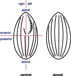
- Antapical: Opposite to the apical region, pertaining to the most posterior point or region of a cell.
- Anterior: The part of the cell in the direction of normal movement; normally the front half of the cell.
- Apical: Pertaining to the most anterior point or region of a cell.
- Dorsal: The side of a cell opposite to the ventral side in cases where the oral region is not at the anterior pole (apex).
- Left: The ciliate’s left side; i.e. the right side of a diagram that displays the ventral surface.
- Posterior: The part of the cell away from the direction of normal movement; normally the antapical half of the cell.
- Right: The ciliate’s right side, i.e. the left side of a diagram that displays the ventral surface.
- Ventral: Generally the region of the cell associated with oral structures, unless the oral region is apical.

- Region opposite to the oral area; generally (but not necessarily) the posterior part of the cell
Aboral
- Polykinetids associated with the oral region in many ciliate taxa
Adoral polykinetids (APk)
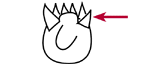
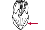
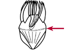
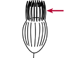
- Polykinetids in strombidiid ciliates that form an incomplete circle or C-shaped band surrounding the oral cavity. Clearly visible in Lugol's fixed material
Adoral polykinetid zone (APZ)
- Some tintinnid ciliates collect various particles (e.g. mineral flakes, coccoliths, diatom frustules) on the outer surface of their lorica
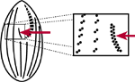
Agglomerated lorica
- Mode of nutrition - synthesising organic compounds using an inorganic source of carbon and energy (e.g. photoautotrophy uses light as an energy source)
Autotrophy
- see kinetosome
Basal body
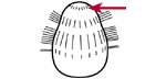
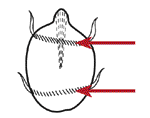
- Distinctive group of cilia arising from short kineties or kinetofragments; often oriented obliquely to the body axis on the anterior-dorsal (or ventral in prostome ciliates) surface. The brosse is used as a diagnostic character for many taxa, but it is not visible on Lugol’s fixed material
Brosse = brush (B)

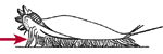
- Distinctly longer somatic cilium (one or more) at or near the posterior pole
Caudal cilium (Cc)
- Some heterotrophic organisms do not digest the chloroplasts from their prey but retain them in specific vacuoles for a certain time. They use the chloroplasts to photosynthesise
Chloroplast retention
- Hair- or whip-like organelle arising from a kinetosomal base and projecting from the surface of the ciliate. Internally it is composed of microtubules typically in a 9+2 arrangement. Normally In ciliates, cilia are numerous and occur on the cell in longitudinal rows, although there are exceptions. Normally visible in Lugol’s fixed material
Cilium (pl. cilia)
- In some genera of the cyclotrichs (Askenasia, Rhabdoaskenasia) the oral area is encircled by this structure. The nature and function of the granules (kinetosomes, extrusomes) is unknown. The circumoral wreath of granules is a diagnostic feature at species level, but cannot be seen in Lugol’s fixed material
Circumoral wreath of granules (CWG)
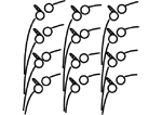
- A polykinetid, functioning as a single unit, often used for locomotion; typical for hypotrichs and stichotrichs, but also used to describe the polykinetids of some cyclotrichs. Clearly visible in Lugol’s fixed material
Cirrus (pl. cirri)
- Outer covering of the cell in which the kinetosomes are embedded
Cortex
- Non-motile, resistant, inactive, dormant stage in the life cycle of some ciliates; generally considered to serve a role in either protection, dispersal, or seasonal dormancy (e.g. over wintering)
Cyst
- Cell-'throat', nonciliated tubular channel leading from the cytostome into the cytoplasm; food vacuoles are typically formed at its inner or distal end; the walls can be strengthened by fibres
Cytopharynx
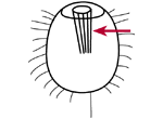
- The funnel-shape arranged bundles of microtubules that may be associated with kinetosomes and form a part of the wall of the cytopharynx in some ciliate groups. May be visible in Lugol’s fixed material
Cytopharyngeal basket (CB)
- Cell mouth; an aperture that may open directly to the exterior or be at the bottom of a depression or cavity, the oral cavity
Cytostome (Cyt)
- Kinetid composed of two kinetosomes plus their cilia and the associated infraciliature
Dikinetid
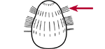
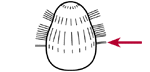
- The posterior end of oligotrichs, below the girdle kinety often has a clear layer that appears expanded in both live and Lugol’s fixed material
Distended cell surface (CS)
- A girdle of short kineties, running parallel to each other, surrounding the cell and bearing cirri; seen in cyclotrichs. Clearly visible in Lugol’s fixed material
Equatorial kinety belt (EKB)
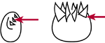
- Type of blade-like oral polykinetids found in strobilidiid and tintinnid ciliates (see external polykinetid zone)
External polykinetid (EPk)
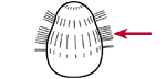
- In strobilidiid and tintinnid ciliates, blade-like oral polykinetids form a full circle around the anterior end of the cell (cf. internal polykinetid zone). Clearly visible in Lugol’s fixed material
External polykinetid zone (EPZ)
- An ejectable organelle, located within the cell; extrusion typically occurs after mechanical or chemical stimuli. Extrusomes may be used for protection or prey capture. Ejected extrusomes are often seen in Lugol’s fixed material
Extrusome (E)
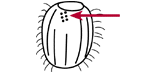
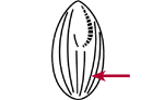
- A kinety in strombidiid ciliates that is wrapped around the cell horizontally, forming one or more whorls, often a characteristic feature at the species level. Although the kinetosomes cannot be seen in Lugol’s fixed material, the position of the kinety is usually visible, and short (2 µm) cilia may be observed
Girdle kinety (G)
- Requiring both energy and organic molecules from the breakdown of organically derived materials
Heterotroph
- Lorica of tintinnid ciliates without agglomerated material on it
Hyaline lorica
- Total assembly of all kinetosomes and their associated structures, located both in somatic and oral regions
Infraciliature
- Type of oral polykinetids found in strobilidiid and tintinnid ciliates (see internal polykinetid zone)
Internal polykinetid (IPk)
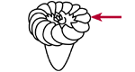
- Polykinetids in strobilidiid and tintinnid ciliates that lie in the oral cavity, within the circle of the external polykinetids; they may be separated from, interdigitated, contiguous or continuous with the external polykinetids. They may be visible in Lugol’s fixed material
Internal polykinetid zone (IPZ)
- One (mono-) or two (di-) kinetosomes = basal bodies plus associated structures; they may be ciliated or unciliated. Not visible in Lugol’s fixed material. The kinetid structure, revealed by transmission electron microscopy, has been extensively used as a diagnostic character for ciliates
Kinetid
- Segments of somatic kineties in the general vicinity of the cytostomal or oral area. Not visible in Lugol’s fixed material
Kinetofragments
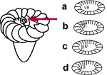
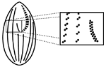
- Within the cortex of the ciliate, the basal body is composed of nine longitudinally oriented, equally spaced sets of three microtubules. If the kinetosome is ciliated, the microtubule triplets are continuous with the paired microtubules of the cilium. Generally not visible in Lugol’s fixed material but can be seen in some preparations using phase or direct interference contrast microscopy. The position, number, and structure of kinetosomes are diagnostic features for ciliates
Kinetosome = basal body
- A row of kinetids, either oral or somatic. These may or may not be ciliated and are often visible in Lugol’s fixed material
Kinety
- A "house", test, envelope, or case, secreted and/or assembled by ciliates, generally fitting the body loosely, opening at one (anterior) or both ends. Clearly visible in Lugol’s fixed material. In planktonic samples, primarily made by tintinnids
Lorica (L)
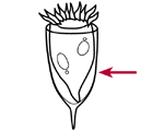
- One of two types of nuclei found in ciliates (cf. micronucleus). Involved in protein synthesis but not in sexual reproduction. Not always visible in Lugol’s fixed material but revealed by DAPI staining (see methods section). The shape, size, number, and position can be diagnostic features for species
Macronucleus (Ma)
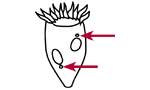
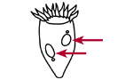
- One of two types of nuclei found in ciliates (cf. macronucleus). Involved in sexual reproduction but not in protein synthesis. Not visible in Lugol’s fixed material but may be revealed by DAPI staining (see methods section). The size, number, and position can be diagnostic features for species
Micronucleus (Mi)
- Heterotrophic, planktonic protists and animals that are between 20 and 200 µm in size
Microzooplankton
- An autotrophic organism that is also able to act as a heterotroph, or a heterotrophic organism that captures chloroplasts from its prey and uses these to photosynthesise (see chloroplast retention)
Mixotroph
- Kinetid composed of one kinetosome
Monokinetid
- Bundle of parallel microtubules, often in a hexagonal arrangement; typically, they are kinetosome-associated; nematodesmata descend into the cytoplasm at a right angle to the cell surface and form the major support of the cytopharyngeal basket of some ciliate groups (e.g. prostomes, some haptorids).
Nematodesma (pl. Nematodesmata)
- referring to the "mouth region" of the ciliate (cf. somatic)
Oral
- Pouch or depression toward the apical end or the ventral surface of the cell; it leads inwardly towards the cytostome and may contain oral polykinetids
Oral cavity
- General term for cilia that are directly associated with the oral apparatus
Oral ciliature (OC)
- Dikinetids that surround the oral region in some prostome ciliates; often they bear long cilia. Not easily seen in Lugol’s fixed samples
Oral dikinetids (OD)
- Long tentacle-like structures found in some prostome species. They originate near the oral dikinetids and surround the oral cavity. Can be seen in Lugol’s fixed material
Oral flaps (OF)
- The subcortical bases of the oral ciliature. Not seen in Lugol’s fixed material
Oral infraciliature
- A row of cilia (kinety) lying along the right side or border of the oral cavity. Not easily seen in Lugol’s fixed material
Paroral kinety (PO)
- Short rows of closely spaced somatic cilia oriented at an oblique angle to the long axis of the body; pectinelles form a circumferential band (= wreath of pectinelles) around the cell, they are found in didiniid ciliates.
Pectinelles (Pec)
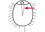
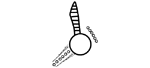
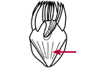
- Elongated protrusion from the posterior end of the tintinnid lorica
Pedicel (P)
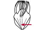
- Posterior cytoplasmic projection, of tintinnid ciliates, which attaches to the inside of the lorica. If the lorica is clear, this can be seen in Lugol’s fixed material
Peduncle (Pd)
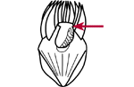
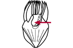
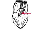
- Cytoplasmic "lip" around the oral cavity. Often visible in Lugol’s fixed material.
Peristomial collar (PC)
- Kinetid composed of more than two kinetosomes plus their cilia and associated structures; found in the somatic region or as part of the oral structures. Can be seen in Lugol’s fixed material
Polykinetid
- Group or series of polykinetids in a close arrangement
Polykinetid zone
- A girdle of short kineties, running parallel to each other, surrounding the anterior half of the cell, bearing cilia or cirri. Seen in cyclotrichs. May be distinguishable from the equatorial kinety belt in Lugol’s samples
Pre-equatorial kinety belt (PKB)
- 1. Trunk-like extension of the anterior end of the cell with the oral area at its base
- 2. Everted cytopharyngeal apparatus (feeding organelle)
Proboscis
- Compound kinetosomal structure in Scuticociliates, non-ciliated and transient. It is located near the posterior base of the paroral kinety, but cannot be seen in Lugol’s fixed material
Scutica (Sc)
- Referring to the "body" of the ciliate (cf. oral)
Somatic
- Cilia or kinetids found anywhere on the surface of the body other than the oral area.
Somatic ciliature
- Row of kinetosomes - mostly longitudinally oriented - on the cell surface, not intimately associated with the oral structures.
Somatic kinety (K)
- A girdle of short kineties, running parallel to each other, surrounding the posterior third of the cell, bearing cilia or cirri. Seen in cyclotrichs. May be distinguishable from the equatorial kinety belt in Lugol’s samples.
Subequatorial kinety belt (SKB)
- Extrusomes situated just below the cortex; they insert into the cortex just above the girdle kinety in strombidiids. Rod-like, 2 - 4 µm in diameter, 5 - 30 µm long, arranged radially in a cone-like formation, often describing the cone-like posterior of oligotrichs. In Lugol’s samples trichites may be seen in the cell, but extruded trichites also occur, extending from just above the girdle kinety.
Trichites (T)
- Somatic kinety, composed of dikinetids, in the posterior, ventral region of most strombidiids; its length and structure are diagnostic, but it is difficult to see in Lugol’s fixed material.
Ventral kinety (Vk)
- A polykinetid in or associated with the oral cavity of strombidiids (see ventral polykinetid zone).
Ventral polykinetid (VPk)
- The band of polykinetids in or associated with the left side of the oral cavity of strombidiids. The polykinetids reduce in size as the zone approaches the cytostome. Most of these can be seen in Lugol’s specimens, if the ventral surface is exposed.
Ventral polykinetid zone (VPZ)
[Printversion]
[Copyright]
[Home]